Anatomy Of Chest Organs : Internal Organs Of The Human Body Anatomical Chart Anatomy ... - The human lungs are a pair of organs which are found on both sides of the chest.
Anatomy Of Chest Organs : Internal Organs Of The Human Body Anatomical Chart Anatomy ... - The human lungs are a pair of organs which are found on both sides of the chest.. The chest contains your heart and lungs; The human lungs are a pair of organs which are found on both sides of the chest. The epidermis is the outermost layer that provides a protective, waterproof seal over the body. A thin, skeletal muscle sitting at the base of the chest, the diaphragm is an unpaired muscle that separates the thorax from the abdomen. Situs inversus (also called situs transversus or oppositus) is a congenital condition in which the major visceral organs are reversed or mirrored from their normal positions.
Anatomy of the chest and stomach, human anatomy, anatomy of the chest and stomach. Where is the sternum found. Computed tomography (ct) of the chest can detect pathology that may not … atlas of ct anatomy of the chest read more » It describes the theatre of events. 12 photos of the anatomy of the chest and stomach.
Anatomy & Physiology 215 > Taylor > Flashcards > Lab 1 ... from classconnection.s3.amazonaws.com The circulatory system does most of its work. This article looks at female body parts and their functions, and it provides an interactive diagram. No need to register, buy now! Anatomy is to physiology as geography is to history: The human thorax includes the thoracic cavity and the thoracic wall. Situs inversus (also called situs transversus or oppositus) is a congenital condition in which the major visceral organs are reversed or mirrored from their normal positions. It is enclosed by the ribs, the vertebral column, and the sternum, or breastbone, and is separated from the abdominal cavity (the body's largest hollow space) by a muscular and membranous partition, the diaphragm. Anatomy of the thoracic (chest) organs.
Find the perfect human chest anatomy stock photo.
The chest or thorax is the region between the neck and diaphragm that encloses organs, such as the heart, lungs, esophagus, trachea, and thoracic diaphragm. Chest the chest consists of bony skeleton of the spine and ribs, chest wall and diaphragm, the mediastinum and great vessels, the airways, lung parenchyma and pulmonary vessels. The human lungs are a pair of organs which are found on both sides of the chest. As you've seen above, the thorax contains more than thoracic arteries, nerves, and lymphatics. The normal arrangement of internal organs is known as situs solitus.although cardiac problems are more common, many people with situs inversus have no medical symptoms or complications resulting from the condition, and. Pathology of the heart, mediastinum, lungs and pleura. The diaphragm forms the upper surface of the abdomen. See chest anatomy stock video clips. Central compartment (mediastinum),… thoracic cage (rib cage). It also contains vital organs and structures, such as the heart, lungs, thymus, trachea, and esophagus. The human thorax includes the thoracic cavity and the thoracic wall. This diagram depicts picture of female reproductive system diagram 1024×1204 with parts and labels. Sternocleidomastoid muscle clavicle and ribs anatomy muscle anatomy chest sternocleidomastoid ribs anatomy chest muscles anatomy thorax rib muscles chest muscles chest anatomy illustration.
System respiratory respiratory organs of human body digestive and respiratory system medical chest internal structure of human body medicine body lungs biology intestines stomach anatomy torso human internal. Among the major organs contained in the thoracic cavity are the heart and lungs. Some run straight through the chest cavity, while others serve nerves and muscles in the cavity which help regulate breathing, the rhythm of the hearth, and the function of various muscles. As you've seen above, the thorax contains more than thoracic arteries, nerves, and lymphatics. Female anatomy includes the external genitals, or the vulva, and the internal reproductive organs.
Thoracic Cavity - Definition & Organs of Chest Cavity ... from biologydictionary.net Anatomy of the chest and stomach, human anatomy, anatomy of the chest and stomach. The chest or thorax is the region between the neck and diaphragm that encloses organs, such as the heart, lungs, esophagus, trachea, and thoracic diaphragm. Chest scan showing a large hydropneumothorax from pleural empyema on the right side of the chest cavity (a is air; Find the perfect human chest anatomy stock photo. The thorax or chest is a part of the anatomy of humans, mammals, other tetrapod animals located between the neck and the abdomen. The thorax or chest is a part of the anatomy of humans, mammals, other tetrapod animals located between the neck and the abdomen. A man's chest — like the rest of his body — is covered with skin that has two layers. The human thorax includes the thoracic cavity and the thoracic wall.
It describes the theatre of events.
Human male muscle body anatomy. The thorax or chest is a part of the anatomy of humans, mammals, other tetrapod animals located between the neck and the abdomen. Posted on july 5, 2015 by admin. At the level of the pelvic bones, the abdomen. Some run straight through the chest cavity, while others serve nerves and muscles in the cavity which help regulate breathing, the rhythm of the hearth, and the function of various muscles. This diagram depicts picture of female reproductive system diagram 1024×1204 with parts and labels. Other minor organs include endocrine glands , lymph ducts, and the esophagus, which simply passes through the thoracic cavity. The chest or thorax is the region between the neck and diaphragm that encloses organs, such as the heart, lungs, esophagus, trachea, and thoracic diaphragm. Internal organs of the human design element, logo. Situs inversus (also called situs transversus or oppositus) is a congenital condition in which the major visceral organs are reversed or mirrored from their normal positions. Anatomy of the chest and stomach, human anatomy, anatomy of the chest and stomach. Central compartment (mediastinum),… thoracic cage (rib cage). When it contracts, the resulting vacuum effect expands and lets you inhale, and then you exhale when this.
This atlas is a comprehensive and affordable. Browse 2,549 female chest anatomy stock photos and images available, or start a new search to explore more stock photos and images. No need to register, buy now! The spongy tissue of the lungs fills with inhaled air thanks to a system of branches referred to as bronchi. It describes the theatre of events.
Internal organs - anatomical model with 4K textures from img2.cgtrader.com At the level of the pelvic bones, the abdomen. The human lungs are a pair of organs which are found on both sides of the chest. This article looks at female body parts and their functions, and it provides an interactive diagram. It describes the theatre of events. Find the perfect chest anatomy stock photo. Human male muscle body anatomy. Anatomy of the chest and stomach, human anatomy, anatomy of the chest and stomach. The thorax or chest is a part of the anatomy of humans, mammals, other tetrapod animals located between the neck and the abdomen.
System respiratory respiratory organs of human body digestive 12.07.2016 · related posts of anatomy of the chest area human anatomy organs female.
When it contracts, the resulting vacuum effect expands and lets you inhale, and then you exhale when this. Internal organs of the human design element, logo. A thin, skeletal muscle sitting at the base of the chest, the diaphragm is an unpaired muscle that separates the thorax from the abdomen. Sternocleidomastoid muscle clavicle and ribs anatomy muscle anatomy chest sternocleidomastoid ribs anatomy chest muscles anatomy thorax rib muscles chest muscles chest anatomy illustration. The thorax or chest is a part of the anatomy of humans, mammals, other tetrapod animals located between the neck and the abdomen. The diaphragm forms the upper surface of the abdomen. The normal arrangement of internal organs is known as situs solitus.although cardiac problems are more common, many people with situs inversus have no medical symptoms or complications resulting from the condition, and. It is enclosed by the ribs, the vertebral column, and the sternum, or breastbone, and is separated from the abdominal cavity (the body's largest hollow space) by a muscular and membranous partition, the diaphragm. Pathology of the heart, mediastinum, lungs and pleura. The human thorax includes the thoracic cavity and the thoracic wall. Anatomy of the chest and stomach, human anatomy, anatomy of the chest and stomach. Learn about each muscle, their locations & functional anatomy. Chest the chest consists of bony skeleton of the spine and ribs, chest wall and diaphragm, the mediastinum and great vessels, the airways, lung parenchyma and pulmonary vessels.
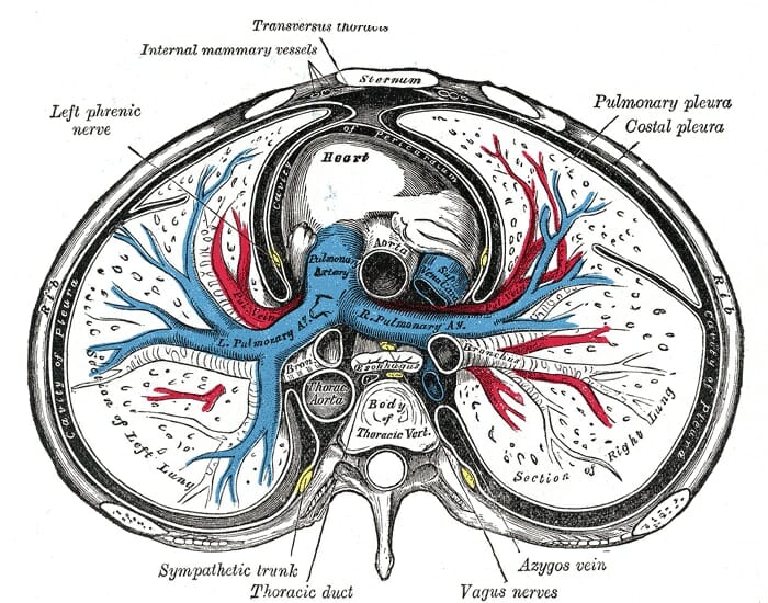
A thin, skeletal muscle sitting at the base of the chest, the diaphragm is an unpaired muscle that separates the thorax from the abdomen anatomy of chest. This image shows some organs of the chest.


0 Komentar